

Windsor Animal Clinic extensively uses X-ray pictures (radiographs) for diagnostic procedures.
Skeleton (orthopedic) X-rays are very helpful in visualizing fractures or bone malformations. Also many congenital defects associated with certain breeds may be detected. These may include bicepital tenosynovitis, shoulder joint dislocation, patellar luxation, ununited anconeal process, elbow joint dislocation, dislocation of the carpus, dislocations of the pelvis, canine hip dysplasia, Perthes’ Disease, cranial cruciate ligament injuries andpatellar luxation. Also diseases such as arthritis, osteochondrosis, panosteitis, hypertrophic osteodystrophy and bone cancers may be detected.
Spinal column injuries can be devastating to an animal because any damage to the delicate spinal cord running through the center of the spine can result in permanent paralysis of portions of the body. Intervertebral disk disease, where the cushion-like disks in between the bones of the spine become deformed, is commonly seen in many breeds. Spinal injuries and diseases are more commonly associated with the branch of medicine entitled neurology.
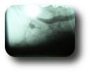
Skull X-rays – Injuries to the skull occur relatively infrequently and are usually associated with blunt trauma, such as an automobile accident.
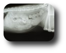
Abdominal X-rays provide an image of the bones and the outlines of a number of internal organs including the liver, stomach, intestines, kidneys, bladder and uterus. This test can be extremely useful for detecting changes in the shape, size or position of organs. Unfortunately, important structures can sometimes blend together on x-rays, so this test does have limitations. For example, a tumor may blend into the background of normal organs because they have the same “opacity,” or shade of gray as the normal tissues. Some foreign objects (such as some plastics) can be invisible on the x-ray. Thus, abdominal x-rays are an excellent “screening test,” but they do not detect all internal problems. In some cases, additional procedures such as ultrasound, endoscopy (scoping), contrast (barium) or dye study or even exploratory surgery are needed to diagnose an intra-abdominal problem.
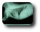
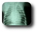
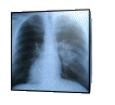
A thoracic (chest) radiograph (X-ray) is a procedure that allows your veterinarian to visualize tissues, organs and bones that lie beneath the skin of the chest cavity. Thoracic radiographs are recommended for any pet with difficultly breathing or with suspicion of heart disease or lung disease. They are also indicated in geriatric patients, and in patients that may have cancer, to evaluate for metastasis (spread). X-rays of the chest should be taken of every animal that has been hit by a car or suffered other types of major trauma because they can reveal many types of injuries to the chest wall, lungs and heart, or other injuries like diaphragmatic hernia. X-rays are also often repeated to monitor progress after treatment or after removing fluid for better visualization of structures. There is no real contraindication to performing this test.
Dental X-rays are also useful when dealing with tooth fractures, abscesses and sinus problems.
![]()